A pioneering global leader in nuclear medicine and molecular imaging
From outstanding healthcare solutions to market excellence: Cyclopharm’s blueprint for growth.
Latest updates
Driving innovation, nurturing growth, enhancing lives.
Cyclopharm, a global radiopharmaceutical industry leader, is synonymous with innovation and excellence in pulmonary care. Under the Cyclomedica banner, we’ve been enabling advancements in groundbreaking lung imaging solutions since 1986 through our flagship product, Technegas®.
Beginning with a commitment to delivering superior patient care and an unwavering focus on improved patient outcomes, our mission extends from laying the foundations to building the deliverables necessary to advance nuclear pulmonology imaging.
As an integral part of Cyclopharm Limited (ASX:CYC), our operations encompass developing state-of-the-art medical equipment, supporting innovative research initiatives into new indications, and cultivating enduring partnerships to expand our product offering. An investment in Cyclopharm is a pledge towards a healthier and more profitable future through our global pursuit of improved patient outcomes.
Downloadable content
Technegas brochureCompelling reasons to invest in Cyclopharm
Surpassing year-on-year sales results
- Sales surge: A 32% record-breaking increase.
- Technegas growth: a notable 4.1% rise.
- Partner distribution: an impressive leap of 124%!
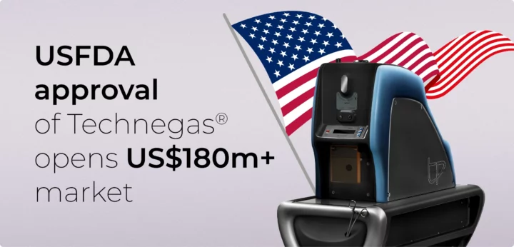
US market debut: Q4 2023 following USFDA approval of Technegas
- $180m USD: Pulmonary Embolism market potential.
- Leading position: USA, hosting half the global nuclear medicine hubs, is Technegas®’ largest potential market.
- Post-approval plan: Elevate Technegas® as the nuclear medicine ventilation imaging gold standard.
- Market expansion: Aiming to double nuclear medicine market share in the USA from 15% to 30%
Established global presence, solid financials with diverse revenue streams
- Global cash flow: Strong earnings from Technegas in 65 nations.
- Diversified revenue: Boosted by third-party distribution.
- Annuity revenue model: The majority of revenues generated from recurring per-patient consumables
- Robust financials: Strong balance sheet to fully fund growth strategy
Upcoming publications: Expanding Technegas clinical application
- Broadening scope: Eyeing new conditions with a potential value of $900m USD.
- Key focus: Pulmonary Embolism, Asthma, COPD, Lung Cancer, Pulmonary Hypertension, Long COVID, Solicosis.
Global regulatory expertise, unique operational footprint & primed for growth
- Compliance success: Regulatory renewals secured under new MDR and MDSAP standards.
- Enviable operational footprint: Leveraging a regulatory, sales and service footprint to diversify customer offering
- Strategic leadership: Board reshaped and ready for upcoming growth
Charting towards a promising future
Cyclomedica’s strategic roadmap unfolds in three stages, designed to cement our position as a world-class provider in molecular imaging. Complemented by the accelerating sales of our partners’ products, Cyclomedica is aiming to emerge as a global billion-dollar enterprise in the forthcoming decade.
Corporate governance
Cyclomedica’s corporate compliance framework encompasses our commitment to sound corporate governance practices by outlining our approach to governance and highlighting the principles that guide our operations.
In the realm of audit and risk management, we acknowledge the inherent potential for unforeseen challenges. At Cyclomedica, we are resolute in our commitment to diligently assess, mitigate, and navigate these risks as they arise. This dedication extends to the thorough monitoring of our operations and adherence to stringent regulatory requirements. In an ever-evolving landscape, our proactive approach to risk management ensures that we remain steadfast in safeguarding our operations against potential disruptions and upholding the highest standards of compliance and accountability.
The possibility exists that Cyclomedica, or our partnering entities, might not consistently adhere to specific legal prerequisites encompassing the development, distribution, and commercialisation of our offerings, along with the holistic operation of our enterprise. This is especially pertinent in areas like competition law, anti-bribery, corruption, and sanctions. Any lapses in maintaining these compliance and legal benchmarks could invite heightened attention and actions from regulatory authorities.
Cyclomedica holds transparency, integrity, and ethical conduct as the cornerstones of our commercial practices and ethical values. We recognise that our actions extend beyond our business operations to influence our stakeholders, partners, and the broader community. With a steadfast commitment to ethical behaviour and responsible business practices, we strive to set industry-leading standards in every facet of our commercial activities. This dedication underscores our pledge to foster trust, uphold fair competition, and maintain the highest ethical standards in all our interactions and endeavours.
Cyclomedica maintains stringent quality systems throughout all our operations and the supply chain to align with industry standards and deliver quality products that are safe, effective, and reliable. Our ongoing commitment to these standards ensures compliance with evolving regulations and scientific insights while underscoring our dedication to transparent and ethical clinical and scientific research.
Share Trading Policy prioritises Cyclomedica’s ethical share trading practices. Insider trading is strictly prohibited, ensuring fairness among shareholders. Clear regulations govern loan-funded share plans, preventing conflicts of interest. We enforce robust reporting procedures to swiftly address suspicious activities, conducting thorough investigations while upholding stringent governance standards. We prioritise transparent, honest disclosure of any material information. Violating the policy carries severe consequences, including legal action. Cyclomedica remains steadfast in its commitment to ethical, compliant share trading, safeguarding the company’s reputation and values.
Past announcements
Subscribe to Cyclopharm’s email alerts service
Whether you are a shareholder or not, you can subscribe to receive all company announcements through our email alerts service.
To join email alerts, simply fill in your details using the form below and nominate which announcements you would like to receive.
Your privacy is important to us. If you no longer want to receive email alerts, use the unsubscribe link. If you are a shareholder and want to change your details or check your holdings, then use the share registry information on the contact us page.


